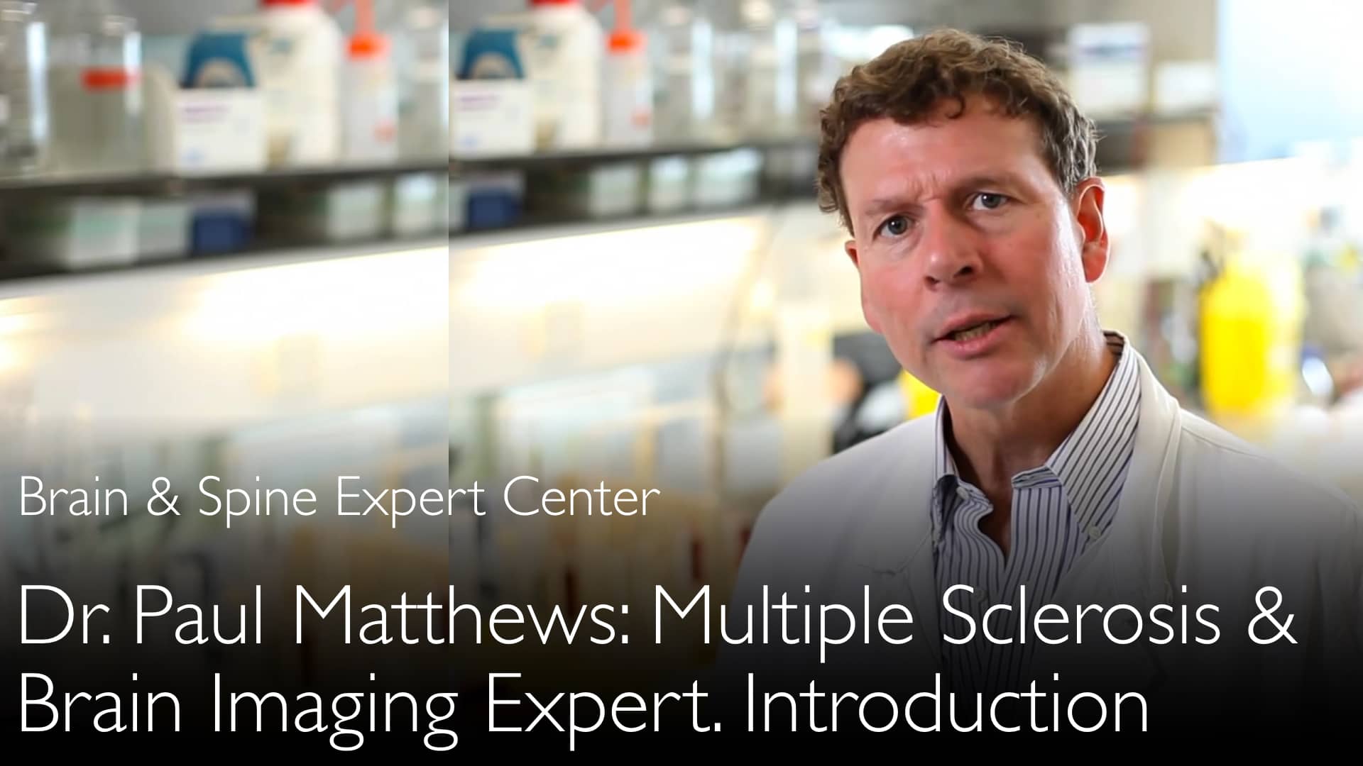
Leading expert in multiple sclerosis and neuroimaging, Dr. Paul Matthews, MD, explains how MRI technology is rapidly evolving. He details advancements in speed, sensitivity, and novel contrast agents. Dr. Paul Matthews, MD, highlights the growing role of quantitative, automated tools for clinical monitoring. He predicts new optical imaging and antibody-based MRI methods will emerge. These innovations will fundamentally improve multiple sclerosis diagnosis and treatment assessment.
Advanced MRI and Future Imaging Technologies for Multiple Sclerosis
Jump To Section
- MRI Speed and Sensitivity Advances
- Novel Antibody-Based Contrast Agents
- Optical Imaging Future Potential
- Quantitative Automated MRI Tools
- Clinical Treatment Criteria Integration
- Full Transcript
MRI Speed and Sensitivity Advances
Dr. Paul Matthews, MD, describes significant progress in MRI examination speed. New multiband techniques have greatly reduced acquisition time for high-quality Diffusion Tensor Imaging. This advanced MRI method provides greater sensitivity for measuring axon bundle density and direction. Dr. Anton Titov, MD, discusses how these fast imaging-based tools are transforming neurological assessment. These improvements allow for more detailed and efficient brain imaging in multiple sclerosis.
Novel Antibody-Based Contrast Agents
Dr. Paul Matthews, MD, highlights emerging antibody-based contrast medications for MRI. These novel agents target inflammation-associated integrins on cerebral vasculature endothelial cells. Dr. Paul Matthews, MD, explains these substances are progressing toward human clinical trials. This represents a major shift from traditional gadolinium-based contrast agents. Dr. Anton Titov, MD, explores how these targeted contrast methods could revolutionize inflammation detection in multiple sclerosis.
Optical Imaging Future Potential
Near-infrared fluorescence methods show promise for multiple sclerosis imaging according to Dr. Paul Matthews, MD. These optical imaging tools provide valuable inflammation data in preclinical models. Dr. Matthews acknowledges the challenges of receiving optical signals from the human brain. The need for exogenous contrast agents presents additional complexity. Despite these hurdles, Dr. Matthews predicts optical imaging will eventually contribute to multiple sclerosis pathology characterization alongside MRI technology.
Quantitative Automated MRI Tools
Dr. Paul Matthews, MD, emphasizes the fundamental shift toward highly quantitative automated imaging tools. These systems generate reproducible data from harmonized acquisition methods. Dr. Paul Matthews, MD, notes this approach mirrors advancements in Alzheimer's disease imaging. Open data sharing accelerates development of new diagnostic tools for multiple sclerosis. Dr. Anton Titov, MD, discusses how commercial online solutions now enable precise brain volume and lesion change measurements.
Clinical Treatment Criteria Integration
Advanced imaging data is becoming integral to clinical treatment decisions in multiple sclerosis. Dr. Paul Matthews, MD, explains how quantitative MRI measures now contribute to therapeutic efficacy monitoring. Practical clinical monitoring tools measure both brain volume change and multiple sclerosis lesion progression. Dr. Matthews describes this as imaging coming full circle. New techniques enhance traditional MRI methods to deliver more actionable clinical information for neurologists treating multiple sclerosis patients.
Full Transcript
Dr. Anton Titov, MD: Where do you see MRI brain imaging technology progressing in the next 5 to 10 years?
Dr. Paul Matthews, MD: More novel imaging methods are increasingly being explored for magnetic resonance imaging. MRI continues to reinvent itself every few years.
First, there are improvements in the speed of examinations by MRI. There is greater sensitivity for measurements, particularly around axon bundle density and direction.
Diffusion tensor imaging MRI improved with the introduction of so-called multiband techniques. This method greatly reduced the time for acquisition of high-quality diffusion tensor data. There are other fast imaging-based tools.
Additional kinds of measures are being explored with novel contrast substances for MRI, although new contrast substances have yet to fully make their way into human use.
For example, antibody-based contrast medications help identify expression of inflammation-associated integrins. These molecules are present on endothelial cells of the cerebral vasculature.
They are increasingly being explored and brought toward human clinical trials by new companies.
Finally, I would emphasize that there is a potential emerging future for additional optical imaging tools. Near-infrared fluorescence methods already provide data in preclinical models that can be related to inflammation.
There's still a long path to go before these will be useful in patients with multiple sclerosis because of the greater complexity of receiving optical signals from the brain.
It is also difficult because of the need for some exogenous contrast agents to be given. But I predict that these tools will eventually contribute to our ability to characterize pathologies such as multiple sclerosis.
Dr. Anton Titov, MD: That's a very small review of the range of imaging tools for better diagnosis of multiple sclerosis.
Dr. Paul Matthews, MD: We continue to move forward. We might expect new brain imaging modalities for multiple sclerosis in the next 10 or 20 years.
The only thing I would add is that the nature of imaging research in multiple sclerosis—MRI, PET, and other imaging technology—is also beginning to fundamentally change.
More and more, the emphasis is on the development of highly quantitative automated tools. They allow very reproducible data to be acquired from increasingly harmonized methods for primary data acquisition.
Secondly, I think there is something we are only beginning to see in multiple sclerosis. This new brain imaging technology is already used for Alzheimer's disease and other major neurodegenerative disorders.
There has been much more open data sharing to allow researchers and doctors to move forward more rapidly in the development of new diagnostic tools. Some of these are becoming commercialized already.
Dr. Anton Titov, MD: There are online solutions for measurement of brain volume change. We can measure multiple sclerosis lesion change. Both of these can contribute to practical clinical monitoring for multiple sclerosis therapeutic efficacy.
Dr. Paul Matthews, MD: Imaging is coming full circle. Brain imaging is getting a new range of techniques, but at the same time, there's a lot of attention paid to making the old MRI methods deliver more information.
Already we are starting to see that information becoming part of clinical treatment criteria in multiple sclerosis.


