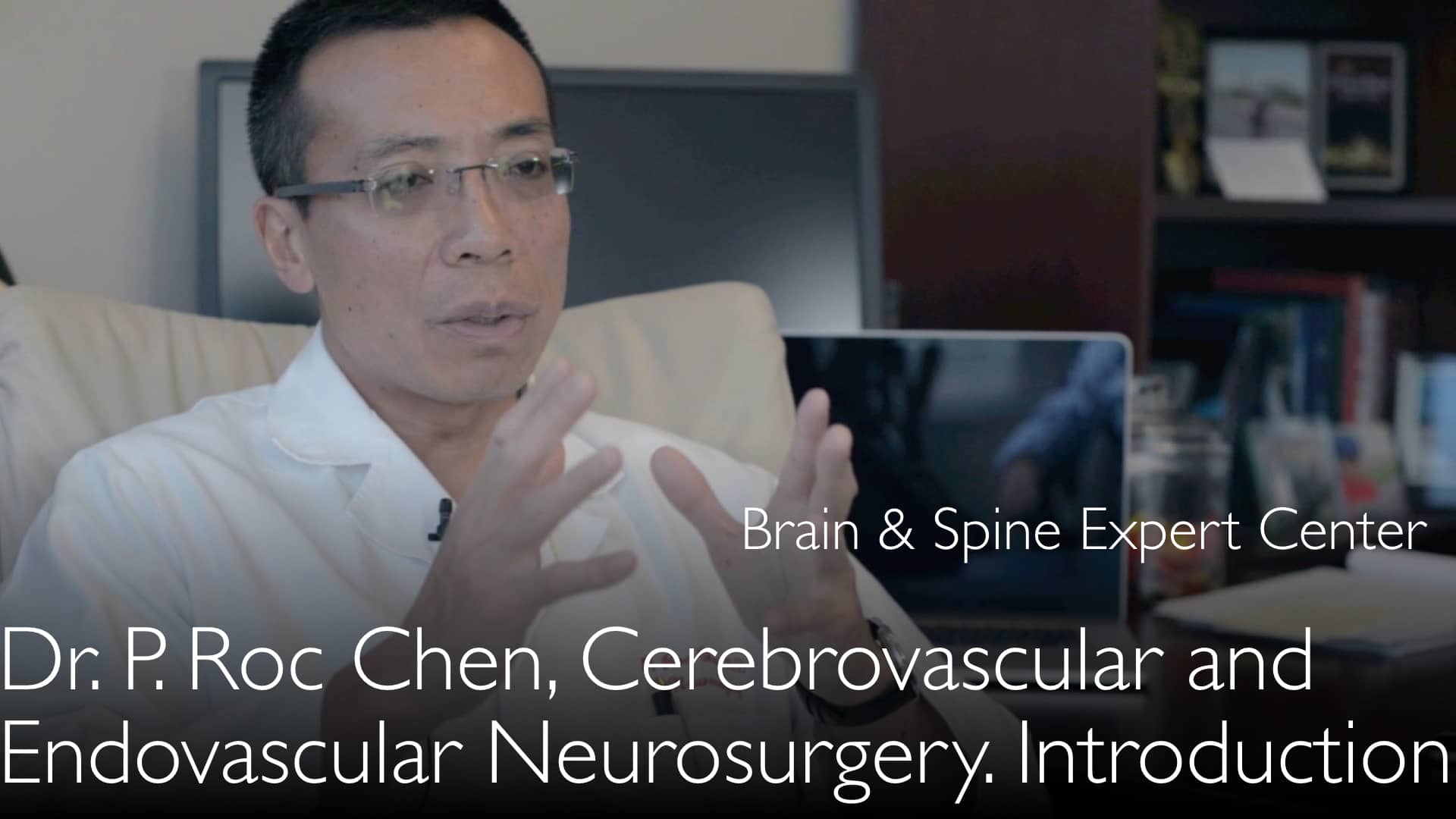
מומחה מוביל בנוירוכירורגיה של כלי דם מוחיים, ד"ר פנג צ'ן, MD, מסביר את תהליך קבלת ההחלטות המורכב בטיפול במלפורמציה עורקית-ורידית (AVM) במוח. הוא מפרט כיצד אסטרטגיית הטיפול תלויה במיקום ה-AVM, גודלו והיסטוריית הקרע, וממליץ על גישה צוותית רב-תחומית המשלבת אמבוליזציה אנדו-וסקולרית וניתוח מוח פתוח להשגת תוצאות מיטביות. ד"ר צ'ן מבהיר כי הסיכון השנתי לדימום ב-AVM שלא נקרע הוא 1-4%, בעוד שהסיכון לדימום חוזר גבוה משמעותית, ועומד על 4-7% לשנה, לאחר קרע ראשוני.
More from Diagnostic Detectives Network

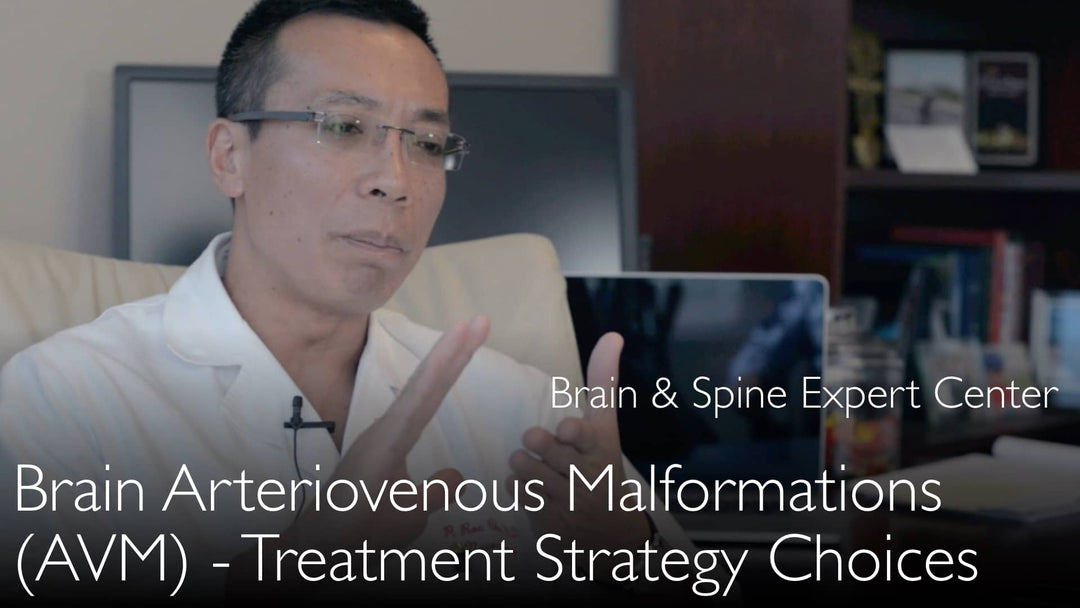
אסטרטגיות טיפול מודרניות למלפורמציות עורקיות-ורידיות במוח (AVMs)
קפיצה לפרק
- מהי מלפורמציה עורקית-ורידית במוח?
- סיכון דימום שנתי של AVM
- סיכוני טיפול לעומת תועלות
- טיפול באנוריזמות נלוות
- טיפול ב-AVM מפוצצת
- גישה טיפולית רב-מקצועית
- תצפית לעומת התערבות
מהי מלפורמציה עורקית-ורידית במוח?
מלפורמציות עורקיות-ורידיות במוח מייצגות קטגוריה של נגעים מוחיים-וסקולריים עם קשרים לא תקינים בין עורקים לוורידים. ד"ר פנג צ'ן, MD, מתאר שלושה סוגים עיקריים. הראשון הוא AVM מולד בזרימה גבוהה, הנוצר במהלך התפתחות העובר וגורם לרשת פלקסיפורמית של קשרים וסקולריים לא תקינים.
הסוג השני הוא פיסטולה עורקית-ורידית דוראלית, המתפתחת לעיתים קרובות במהלך חיי המטופל, ככל הנראה עקב פקקת ורידית. הסוג השלישי הוא מלפורמציה מערתית, או קוורנומה, שאינה נראית בהדמיה וסקולרית סטנדרטית אך ניתן לראותה בסריקות MRI ו-CT, וגורמת לעיתים קרובות לדימומים קטנים.
סיכון דימום שנתי של AVM
סיכון הפיצוץ של מלפורמציה עורקית-ורידית במוח וגרימת דימום הוא גורם קריטי בהחלטות טיפול. לפי ד"ר פנג צ'ן, MD, הסיכון השנתי לדימום עבור מלפורמציה עורקית-ורידית במוח שלא התפוצצה הוא בדרך כלל בין 1% ל-4%. סיכון זה אינו אחיד ותלוי heavily במאפייני ה-AVM הספציפיים.
ד"ר פנג צ'ן, MD, מסביר כי סיכון הפיצוץ תלוי בגודל ובגיאומטריה של התעלות הווסקולריות בתוך המלפורמציה. הוא מציין ש-AVMs גדולות מאוד, המסווגות כדרגה 4 או 5 בסולם Spetzler-Martin, עשויות למעשה להיות בעלות סיכון פיצוץ נמוך יותר ממה שסברו בעבר, פחות מ-1% לשנה, במיוחד כאשר סיכון מאנוריזמות נלוות מחושב בנפרד.
סיכוני טיפול לעומת תועלות
שקילת סיכוני הטיפול מול הסיכון הטבעי לדימום היא אבן היסוד בטיפול במלפורמציות עורקיות-ורידיות במוח. ד"ר פנג צ'ן, MD, מדגיש כי הסיכונים מטיפול כירורגי או אנדו-וסקולרי אגרסיבי ב-AVMs גדולות יכולים להיות לעיתים גבוהים יותר מסיכון פיצוץ ספונטני. הבנה זו הובילה לגישה שמרנית יותר במקרים מורכבים מסוימים.
לעומת זאת, ד"ר צ'ן מציין ש-AVMs קטנות יותר נושאות לעיתים קרובות סיכון דימום יחסי גבוה יותר. כאשר אלו ממוקמות באזורים לא אלוקוונטיים, או פחות קריטיים, של המוח, ניתן לבצע טיפול בצורה מאוד בטוחה. בתרחישים אלו, התועלת בחיסול סיכון הדימום העתידי גוברת על סיכוני ההליך, מה שהופך את ההתערבות למסלול הפעולה המועדף.
טיפול באנוריזמות נלוות
מטופלים עם מלפורמציות עורקיות-ורידיות במוח יכולים לפתח אנוריזמות נלוות עקב שנים של זרימת דם לא תקינה בלחץ גבוה. ד"ר פנג צ'ן, MD, מדגיש שאנוריזמות מוחיות אלו עצמן נושאות סיכון דימום מוגבר משמעותית. סיבוך זה דורש תשומת לב ספציפית במהלך הערכת המטופל.
אסטרטגיית הטיפול מתמקדת לעיתים קרובות בטיפול באנוריזמה תחילה. ד"ר פנג צ'ן, MD, מציין שאנוריזמות אלו יכולות frequently להיות מטופלות successfully בשיטות אנדו-וסקולריות, שפחות פולשניות מניתוח פתוח. טיפול נפרד באנוריזמה הוא צעד crucial בהפחתת הסיכון הכללי לדימום עבור המטופל.
טיפול ב-AVM מפוצצת
פרדיגמת הטיפול משתנה significantly לאחר שמלפורמציה עורקית-ורידית במוח התפוצצה. ד"ר פנג צ'ן, MD, קובע כי הסיכון לדימום חוזר גבוה much, ונע between 4% ל-7% לשנה. סיכון מוגבר זה means שמטופלים עם AVM מפוצצת יזדקקו generally לצורה כלשהי של טיפול למניעת דימום נוסף.
עקרונות הטיפול ב-AVM מפוצצת involve אותם modalities—ניתוח, אמבוליזציה, ורדיו-כירורגיה. עם זאת, הדחיפות וההכרח להשמדה complete גדולים more. תהליך קבלת ההחלטות remains complex, במיוחד אם ה-AVM ממוקמת באזור אלוקוונטי של המוח, הדורש תכנון careful על ידי צוות רב-מקצועי.
גישה טיפולית רב-מקצועית
טיפול מודרני במלפורמציות עורקיות-ורידיות במוח demands גישה collaborative, רב-מקצועית. ד"ר פנג צ'ן, MD, advocate strongly שטיפול should not rely on שיטה single. He insists שהתוצאות best מושגות through שילוב של אמבוליזציה אנדו-וסקולרית וניתוח מוח פתוח, המותאמים לאנטומיה של המטופל individual.
תהליך זה begins with תוכנית טיפול meticulously detailed המנוסחת על ידי צוות including neurosurgeons, מומחים אנדו-וסקולריים, ו-experts רדיו-כירורגיה. המטרה היא להשמיד completely את ה-AVM while לשמר function, particularly כאשר היא ממוקמת near אזורי מוח critical. ד"ר צ'ן warns against טיפול incomplete, "half-way", which can be ineffective and leave the patient at risk.
תצפית לעומת התערבות
ההחלטה between watchful waiting and התערבות active is nuanced. ד"ר פנג צ'ן, MD, explains that observation may be a valid strategy for large, unruptured AVMs in eloquent locations where treatment risks are prohibitively high. This is especially true given the potentially lower annual bleeding rates for these complex lesions.
However, for younger patients or those with AVMs in safer locations, intervention is often recommended. The cumulative lifetime risk of hemorrhage justifies treatment. Dr. Chen concludes that a thorough assessment by a specialized team is essential before deciding on any management path, ensuring the chosen strategy aligns with the patient's specific risk profile and anatomy.
תמליל מלא
טיפול במלפורמציות עורקיות-ורידיות במוח יכול להיות very complex. תצפית, ניתוח מוח פתוח, או אמבוליזציה אנדו-וסקולרית הם שלוש שיטות טיפול ל-AVMs מוחיות. They can be used together or sequentially.
אסטרטגיית טיפול במלפורמציות עורקיות-ורידיות במוח depends on location and type of AVM. Probability of cerebral arteriovenous malformation hemorrhage is higher with AVMs that contain a brain aneurysm.
ד"ר פנג צ'ן, MD: מלפורמציה עורקית-ורידית היא אינדיקציה לניתוח מוח פתוח ולטיפול אנדו-וסקולרי. סיכון דימום AVM depends on previous history of hemorrhage and AVM size and shape. מלפורמציות עורקיות-ורידיות תוך-גולגולתיות בדרגת Spetzler Martin AVM 4 is lower than usually reported. מיקומי אזורי מוח אלוקוונטיים של AVM have to be assessed for watchful waiting observation.
מהם ניואנסי הטיפול במלפורמציה עורקית-ורידית במוח? מהן שיטות מודרניות לטיפול במלפורמציות עורקיות-ורידיות מוחיות?
ד"ר פנג צ'ן, MD: מלפורמציות עורקיות-ורידיות במוח (brain AVM) הן קטגוריה גדולה של נגעים מוחיים-וסקולריים. הגדרה רחבה של מלפורמציות עורקיות-ורידיות היא זו: המוח includes מלפורמציות עורקיות-ורידיות בזרימה גבוהה ופיסטולות עורקיות-ורידיות דוראליות. מלפורמציות עורקיות-ורידיות מוחיות are usually congenital. They form during 4 to 8 weeks of fetal development, when vascular structures develop in the fetus.
Wrong development of cerebral blood vessels results in plexiform web of incorrectly formed connections between arteries and veins. Second type of vascular malformation in the brain is called brain dural arteriovenous fistula (BDAVF). This malformation often forms during patient's lifetime. We do not know the reasons why brain dural arteriovenous fistulas develop. They could be a result of venous thrombosis in the brain, but we do not really know.
Third type of brain arteriovenous malformation is cavernous malformation (cavernous angioma). They are not visible on vascular imaging, but brain MRI and CT scans can show cavernous malformations well. Brain cavernous malformations can cause brain hemorrhage (bleeding) on a small scale. So these are three types of brain arteriovenous malformations.
High-flow brain arteriovenous malformations have a risk of bleeding into the brain. The risk of brain hemorrhage from unruptured cerebral arteriovenous malformations is a controversial issue. Probability of rupture of brain arteriovenous malformation has been studied. However, we do not know with certainty how big is the risk of bleeding.
I think that the risk of rupture of any unruptured brain arteriovenous malformation is 1% to 4% per each year. This risk of bleeding depends on the size of arteriovenous malformation. The risk of brain arteriovenous malformation rupture also depends on geometry of vascular channels inside the arteriovenous malformation.
Very large brain arteriovenous malformations are called "Spetzler Martin AVM grade 4" or "Spetzler Martin AVM grade 5". Risk of rupture of large brain AVMs decreases if we deduct the risk of rupture of brain aneurysms. Brain aneurysms can sometimes co-exist with brain arteriovenous malformations.
The risk of rupture and bleeding of these large brain arteriovenous malformations (Spetzler Martin grade 4 and 5) is much lower than we previously thought. Risk of rupture of brain arteriovenous malformation is about 1% per each year, or it is less than 1% per each year.
We previously treated large brain arteriovenous malformations very aggressively. But risks from surgical and endovascular treatment of large brain AVMs are also very high. Risks of treatment could be higher than risks of spontaneous aneurysm rupture. Therefore, we currently do not aggressively treat very large brain arteriovenous malformations.
Sometimes patients with brain AVMs also have brain aneurysms. Aneurysms form in such patients because of many years of high pressure blood flow through brain vessels. Brain aneurysms in patients with cerebral arteriovenous malformations carry increased risk of bleeding from the aneurysm. We can often treat brain aneurysms in such patients by endovascular methods.
Approximately two thirds of patients with large cerebral arteriovenous malformations may have seizures. We can successfully treat seizures in these patients too. The risk of bleeding from rupture of cerebral arteriovenous malformation is higher in those patients who have brain arteriovenous malformations of smaller size.
Sometimes brain arteriovenous malformation is located in the area of brain that does not contain very important functional area around AVM. Then we can treat brain arteriovenous malformation very safely. It is better to treat such cerebral arteriovenous malformations because the risk of their rupture is higher than the risk of surgical or endovascular treatment.
Risk of brain AVM rupture is higher in younger patients. Particularly younger patients can benefit from open brain surgery or endovascular embolization procedure. Combination of both treatments methods can often remove cerebral arteriovenous malformation completely.
Stereotactic radiosurgery ("gamma knife") can be used to treat brain arteriovenous malformation in areas of the brain that are very difficult to reach or where surgery can damage brain function. But it is most important to carefully and fully assess the situation of the patient with brain arteriovenous malformation before deciding about any method of treatment or observation of brain arteriovenous malformation.
טיפול חלקי ולא מלא בחולים עם AVM מוחי לא מפוצץ אינו מומלץ. עדיף להשתמש בשילוב של אמבוליזציה אנדווסקולרית וניתוח מוח פתוח. אין להשתמש בשיטת טיפול אחת בלבד. על הרופאים לנקוט בגישה רב-מקצועית (multidisciplinary team approach).
על המנתחים להכין בקפידה תוכנית טיפול מפורטת למלפורמציה עורקית-ורידית מוחית לפני תחילת כל שיטת טיפול. במקרים של מלפורמציה עורקית-ורידית מוחית מפוצצת, הסיכון לפיצוץ חוזר גבוה בהרבה. הסיכון לדימום מוחי חוזר הוא 4% לשנה עד 7% לשנה. לכן אנו יודעים כי חולים עם AVM מוחי מפוצץ בדרך כלל זקוקים לטיפול.
עקרון הטיפול במלפורמציות עורקיות-ורידיות מוחיות מפוצצות זהה לזה של טיפול ב-AVM מוחי לא מפוצץ. לעיתים ה-AVM המוחי ממוקם באזור פחות חשוב במוח. ניתן להשתמש בניתוח מוח פתוח.
לעיתים המלפורמציה העורקית-ורידית המוחית ממוקמת באזור במוח בעל קליפה תפקודית חשובה. אז ההחלטה על הטיפול מורכבת וקשה. נוירוכירורגים, מומחים לנוירוכירורגיה אנדווסקולרית ומומחים לרדיו-כירורגיה צריכים להחליט כיצד לחסל מלפורמציות עורקיות-ורידיות מוחיות וכיצד לטפל בחולה ללא פגיעה באזורים תפקודיים חשובים במוח.
זהו הרמה הנוכחית של הגישה שלנו לטיפול במלפורמציות עורקיות-ורידיות מוחיות. טיפול במלפורמציות עורקיות-ורידיות מוחיות: מעקב או התערבות? אמבוליזציה אנדווסקולרית או ניתוח מוח פתוח? התקדמויות בטיפול ב-AVM.