מומחה מוביל ברפואת אם ועובר, ד"ר איב ויל, MD, מסביר מדוע בדיקות צוואר הרחם הדיגיטליות מיושנות ומסוכנות. הוא מפרט כיצד אולטרסאונד וגינלי מספק מדידה אובייקטיבית מעולה של אורך צוואר הרחם לחיזוי סיכון ללידה מוקדמת. ד"ר ויל דן גם בתפקיד הקריטי של MRI (דימות תהודה מגנטית) באבחון מצבי שליה מורכבים כמו פלצנטה אקרטה, תוך מתן פרטים אנטומיים חסרי תקדים לניהול הריון בטוח יותר.


טיפול טרום לידתי מודרני: החלפת בדיקות ידניות באולטרסאונד ו-MRI
קפיצה לפרק
- סכנות וחוסר הדיוק של בדיקות צוואר הרחם הידניות
- אולטרסאונד וגינלי לחיזוק לידה מוקדמת
- הבנת הערך החיזוי בהערכת צוואר הרחם
- MRI לאנטומיה מפורטת של העובר והשליה
- אבחון ספקטרום חדירת השליה באמצעות MRI
- עתיד הדימות התפקודי בהריון
- תמליל מלא
סכנות וחוסר הדיוק של בדיקות צוואר הרחם הידניות
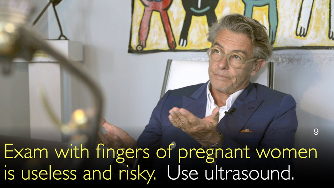
ד"ר איב ויל, MD, מציין כי בדיקה ידנית של צוואר הרחם היא פרקטיקה בת מאות שנים שיש לזנוח במהלך ההריון. הוא מדגיש כי שיטה זו חסרת תועלת מחוץ לשלב הלידה הפעילה. ההליך רצוף אי-דיוקים, ומפיק תוצאות חיוביות ושליליות כוזבות.
באופן קריטי יותר, ד"ר איב ויל, MD, מדגיש את סיכון הזיהום המשמעותי הקשור בבדיקה ידנית. הוא מסביר כי צוואר רחם ארוך לכאורה עשוי להיות פתוח פנימית, והכנסת אצבעות יכולה ליישב חיידקים בקרומים. יתרה מכך, צוואר רחם עשוי להרגיש קצר במגע אך להימדד כאורך באולטרסאונד, מה שמדגים את חוסר האמינות הבסיסי של השיטה.
אולטרסאונד וגינלי לחיזוק לידה מוקדמת
ד"ר איב ויל, MD, תומך באולטרסאונד וגינלי כתקן הזהב האובייקטיבי להערכת סיכון ללידה מוקדמת. טכניקת דימות זו מודדת במדויק את אורך צוואר הרחם ויכולה לזהות שינויים קריטיים, כגון משפך, שבו הפתיחה הפנימית של צוואר הרחם מתחילה להתרחב. אולטרסאונד מספק הערכה ויזואלית ברורה שלא ניתן להשיג בבדיקה גופנית.
ד"ר אנטון טיטוב, MD, דן בהתקדמות זו עם ד"ר ויל, שהעיר על חשיבותה המיוחדת להריונות בסיכון גבוה. עבור נשים עם היסטוריה של לידה מוקדמת או אלו הנושאות תאומים או שלישיות, מדידה ארוכה של צוואר הרחם באולטרסאונד מספקת הרגעה עצומה. נתונים אובייקטיביים אלה הוכחו כעדיפים על בדיקה ידנית במחקרים קליניים רבים.
הבנת הערך החיזוי בהערכת צוואר הרחם
חוזק מרכזי של אולטרסאונד, כפי שהוסבר על ידי ד"ר איב ויל, MD, הוא הערך החיזוי השלילי המעולה שלו. כאשר אורך צוואר הרחם ארוך, הסבירות ללידה מוקדמת נמוכה מאוד. זה מספק שקט נפשי קריטי למטופלות ולקלינאים המטפלים בתרחישים בסיכון גבוה.
ד"ר ויל מבהיר כי בעוד שאולטרסאונד עדיף בהרבה על בדיקה ידנית, צוואר רחם קצר בלבד אינו מבטיח תמיד לידה מוקדמת. הערך החיזוי החיובי אינו חזק באותה מידה. זה הוביל למחקר על בדיקות אבחון משלימות, כמו פיברונקטין עוברי, כדי לשפר את הדיוק בחיזוי אילו נשים עם צוואר רחם קצר אכן יילדו מוקדם.
MRI לאנטומיה מפורטת של העובר והשליה
במהלך הריאיון עם ד"ר אנטון טיטוב, MD, ד"ר איב ויל, MD, פירט את שני היישומים העיקריים של MRI בהריון: מורפולוגיה ותפקוד. לפרטים אנטומיים, MRI הוא כלי מדויק ביותר, במיוחד לדימות מוח העובר בשליש השלישי. הבהירות שלו טובה בהרבה מאולטרסאונד בהריון המאוחר.
ד"ר איב ויל, MD, מציין כי MRI מספק את הערך החיזוי השלילי הטוב ביותר לשלילת מומים מוחיים, במיוחד לאחר חשד לזיהום עוברי. בעוד שאולטרסאונד עדיין עדיף על MRI בהערכת תפקוד השליה באמצעות טכנולוגיית דופלר, MRI מפתח במהירות את יכולותיו לדימות תפקודי של המוח, השליה ואיברים אחרים.
אבחון ספקטרום חדירת השליה באמצעות MRI
ד"ר איב ויל, MD, מזהה את MRI ככלי מושלם לאבחון הפרעות ספקטרום חדירת השליה. מצב מסכן חיים זה מתרחש כאשר השליה פולשת עמוק מדי לדופן הרחם, ללא מישור הפרדה נורמלי. הספקטרום כולל שליה אקרטה, אינקרטה והחמורה ביותר, פרקרטה, שבה השליה פולשת דרך דופן הרחם.
ד"ר איב ויל, MD, מסביר כי MRI חשוב במיוחד כאשר השליה ממוקמת על הדופן האחורית של הרחם, שם אולטרסאונד פחות יעיל. הסריקה יכולה להראות בבירור את עומק הפלישה ולהעריך מעורבות של כלי דם אימהיים סמוכים. מידע אנטומי מפורט זה הוא גורם פרוגנוסטי חיוני לתכנון כירורגי ויכול גם לשלול definitively את האבחנה כדי להקל על חרדת המטופלת.
עתיד הדימות התפקודי בהריון
הדיון בין ד"ר אנטון טיטוב, MD, וד"ר איב ויל, MD, הביט לעתיד הדימות הטרום לידתי. בעוד שיישומי האנטומיה של MRI מבוססים היטב, התפקיד התפקודי שלו עדיין מתפתח. מחקר מתמשך להשתמש ב-MRI לחקור תפקוד מוחי ופרפוזיה שלייתית מפורטת.
ד"ר ויל מסיים כי שילוב אמצעי הדימות המתקדמים הללו מייצג את תקן הטיפול החדש. החלפת בדיקות ידניות סובייקטיביות באולטרסאונד ו-MRI אובייקטיביים מובילה לאבחנות מדויקות יותר, שכבות סיכון טובות יותר, ובסופו של דבר, תוצאות בטוחות יותר עבור האם והילד.
תמליל מלא
ד"ר אנטון טיטוב, MD: אתה גם מומחה במניעת לידה מוקדמת. פרסמת מחקר חשוב באמצעות בדיקת אולטרסאונד וגינלי למדידת אורך צוואר הרחם. זה יכול לחזות את הסיכון לקריעה מוקדמת של הקרומים, לידה מוקדמת, וכמה סיבוכים חמורים עבור האם והילד. אנא דון בעבודה שלך במניעת לידה מוקדמת.
ד"ר איב ויל, MD: נכון. מיילדות במשך מאות שנים, אלפי שנים, מבוססת על בדיקה ידנית של צוואר הרחם, שהיא הדבר הגרוע ביותר שיכולנו להעלות על הדעת. זה חסר תועלת במהלך ההריון. זה פחות בשימוש בעולם האנגלו-סקסי, וזה דבר טוב מאוד.
אני חושב שאף אישה בהריון לא צריכה להיבדק ידנית במהלך ההריון. היתרון היחיד של בדיקה ידנית של צוואר הרחם הוא כאשר אישה בלידה. מחוץ למצב זה, זה מלא במידע חיובי ושלילי כוזב.
בדיקה ידנית של צוואר הרחם מגבירה את סיכון הזיהום. זה מגביר את סיכון הזיהום כי צוואר רחם ארוך יכול להיות פתוח entirely ומיושב על ידי הקרומים מבפנים, והבדיקה הידנית שלך תהיה נורמלית.
צוואר רחם יכול להרגיש קצר עם האצבעות שלך ולהיות ארוך באולטרסאונד עם אוסטיום פנימי שהוא מאוד גבוה. וברגע שאישה ילדה פעם אחת בחייה, צוואר הרחם never סגור שוב. לכן, לומר ש"אה כן, אני לא יכול להכניס את האצבע שלי", certainly אל תעשה את זה. ואם אתה עושה את זה, זה לא מוכיח anything.
אז אולטרסאונד emerged as הבדיקה האובייקטיבית ביותר לגבי סיכון ללידה מוקדמת. צוואר רחם קצר או צוואר רחם פתוח, זה called משפך מבפנים, although הוא ארוך, יכול להיות בסיכון גבוה. אולטרסאונד מאוד מדויק for that.
Also, and most importantly, במצב בסיכון גבוה, נשים עם היסטוריה קודמת של לידה מוקדמת, נשים עם תאומים או שלישיות, אם צוואר הרחם באולטרסאונד ארוך, then הערך החיזוי השלילי של לידה מוקדמת extremely גבוה.
So אולטרסאונד proven in many clinical studies to be superior to בדיקה ידנית, and to have an excellent negative predictive value. It's only when a cervix is short and there are contractions. If the cervix is short with ultrasound, although it's better than digital examination, the positive predictive value is still not that high.
So people have worked on a complementary diagnostic test for that. Fibronectin and other things to try and improve the positive predictive value for premature labor. But the negative predictive value of ultrasound is excellent.
So ultrasound in obstetrics has become a clinical tool. Even if it's an oxymoron because it's a device, it's not clinical. It is the best clinical examination is vaginal ultrasound in obstetrics.
Dr. Anton Titov, MD: Thank you, that's very important information. You're also a leading expert on MRI in the diagnosis of problems during pregnancy, both for the unborn mother and developing child. An MRI of the placenta can identify important risk factors for the developing fetus, for the mother, and for the future child. How can MRI be used for the diagnosing of problems in the placenta? And what is the future for MRI use in diagnosing problems during pregnancy?
Dr. Yves Ville, MD: Schematically, there are two kinds of expectations for MRI. One expectation is about morphology. The other one is about function. For morphology, MRI is an extremely accurate tool. Both for the fetal anatomy and especially the brain, MRI is unbeatable, much better than ultrasound, especially late in the pregnancy.
And if you look at function, today ultrasound is much more accurate than MRI in predicting functional problems with the placenta, for example, by using Doppler, umbilical and fetal Doppler. MRI is currently developing in exploring the function of the brain, function of certain organs, and function of the placenta.
But to date, MRI has not achieved a clinical level of performance. Although if you look at, again, the anatomy, the anatomy of the brain, the fetal brain in a high-risk situation, we mentioned infections earlier on, MRI has the best negative predictive value. Clearly.
Now, MRI of the placenta is extremely useful and accurate if you suspect placenta accreta or if you have risk factors for placenta accreta. So, placenta accreta is a placenta inserted not on the surface of the uterus with a cleavage plane, represented by the endometrium. It is going through the endometrium, at a different level of penetration of the uterus, sometimes down to the serosa.
So you got placenta accreta, placenta increta, placenta percreta. Especially when the placenta is posterior, ultrasound is not the best tool for diagnosing placenta accreta. But MRI is pristine to show you either placenta accreta, increta, or percreta.
You can look at the invasion of the nearby vessels in the pregnant woman, which is a very important prognostic factor for the management and mainly to exclude placenta accreta, actually. You can say, "No, don't worry, this placenta looks odd on ultrasound, but there is no placenta accreta."