
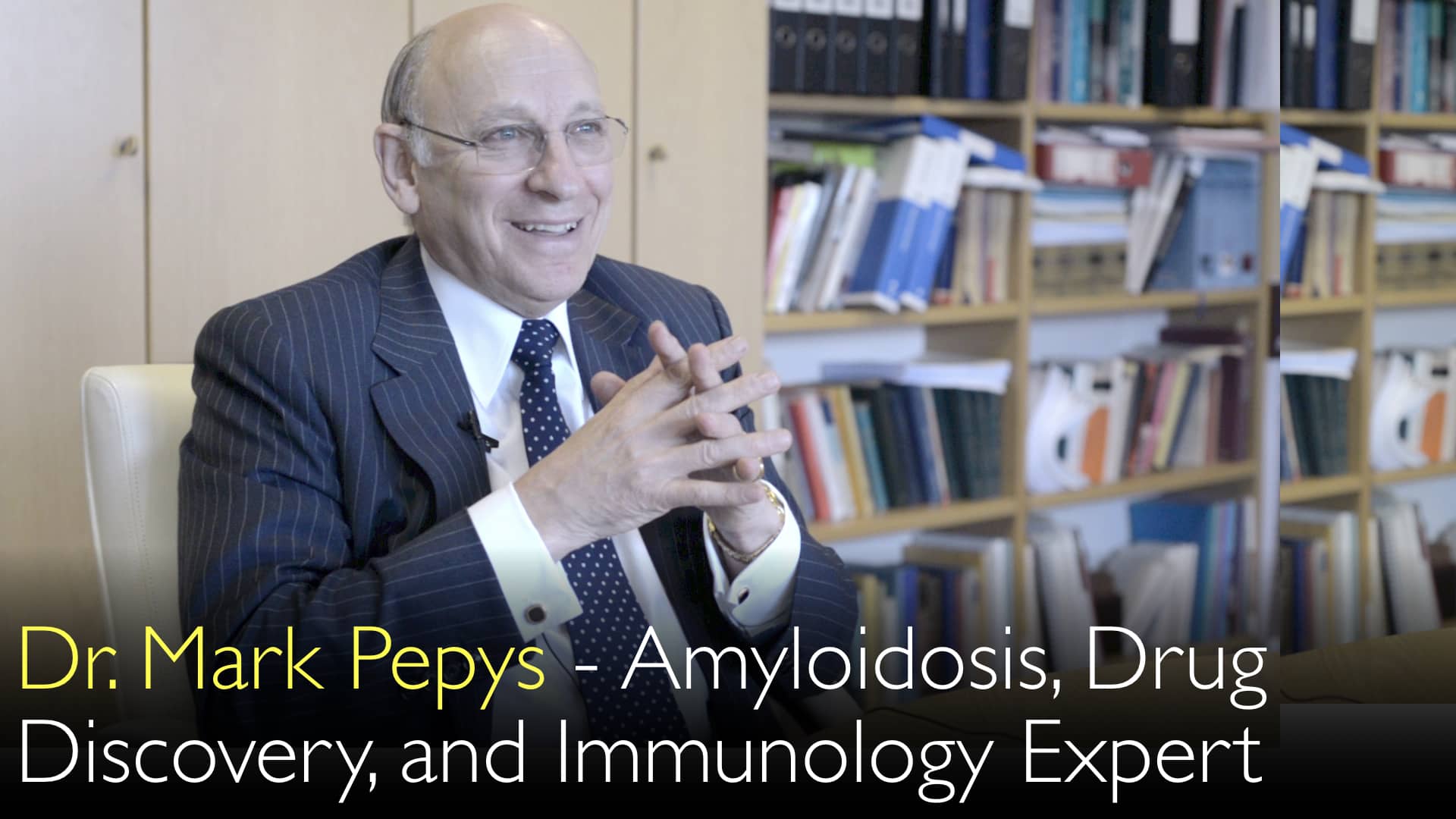
מומחה מוביל באבחון וטיפול באמילואידוזיס, ד"ר מארק פפס, MD, מסביר את האתגרים האבחוניים של מחלה נדירה זו. תסמיני אמילואידוזיס מחקים מחלות רבות אחרות. רופאים לעיתים קרובות אינם שוקלים אמילואידוזיס בתחילה. הבדיקה האבחונית הנכונה היא ביופסיה. אמילואידוזיס קשור להפרעות בתאי פלזמה כמו מיאלומה נפוצה. המחלה כרוכה בחלבונים מסיסים בדרך כלל שמתקפלים בצורה לא נכונה לסיבי אמילואיד בלתי מסיסים. תעלומה רפואית מרכזית היא מדוע המקרופאג'ים של הגוף אינם מצליחים לפנות משקעי חלבון חריגים אלה. ד"ר מארק פפס, MD, דן במחקרו על התערבויות טיפוליות לתיקון כשל זה.
More from All
More from Diagnostic Detectives Network

הבנת עמילואידוזיס: גורמים, אתגרים באבחון ומחקרי טיפול
קפיצה לפרק
- אתגרים באבחון עמילואידוזיס
- קשר למיאלומה נפוצה
- תהליך יצירת העמילואיד
- תעלומת פינוי הגוף
- כיווני מחקר טיפולי
- תמלול מלא
אתגרים באבחון עמילואידוזיס
אבחון עמילואידוזיס מציב קשיים קליניים משמעותיים לפי ד"ר מארק פפיס, MD. ביטויי המחלה מגוונים ביותר ויכולים לחקות מצבים שכיחים רבים אחרים. שונות זו意味着meansרופאים לרוב אינם מחשיבים עמילואידוזיס באבחנה המבדלת הראשונית שלהם.
ד"ר אנטון טיטוב, MD, דן עם ד"ר פפיס כיצד התעלמות אבחונית זו מובילה לעיכובים בהזמנת הבדיקות הנכונות. הבדיקה האבחונית המוחלטת לעמילואידוזיס היא ביופסיית רקמה הנבדקת תחת מיקרוסקופ. ד"ר מארק פפיס, MD, מדגיש כי מקרים חשודים יש להפנות למרכזים מומחים להדמיה מתקדמת ולהליכים אבחוניים.
קשר למיאלומה נפוצה
לעמילואידוזיס קשרים חשובים להפרעות בתאי פלזמה כמו מיאלומה נפוצה. ד"ר מארק פפיס, MD, מסביר שמיאלומה נפוצה מייצגת מחלה ממאירה סרטנית within the spectrum of monoclonal gammopathy. אותם תאי פלזמה שהופכים לסרטניים במיאלומה יכולים גם לגרום לעמילואידוזיס מבלי להיות ממאירים בעצמם.
ד"ר אנטון טיטוב, MD, וד"ר פפיס מציינים שתאים non-cancerous אלה אינם מתרבים ללא שליטה או פולשים לאיברים. במקום זאת, הם מייצרים חלבונים לא תקינים היוצרים משקעי עמילואיד. הסוג הנפוץ ביותר של עמילואידוזיס מערכתי נגרם על ידי חלבונים these plasma cell-derived, creating diagnostic confusion between the two conditions.
תהליך יצירת העמילואיד
התהליך הבסיסי בעמילואידוזיס involves protein misfolding and aggregation. ד"ר מארק פפיס, MD, משתמש באנלוגיה of egg white transformation להסביר concept זה. חלבונים מסיסים normally become insoluble through physical state changes, similar to how liquid egg white turns solid and opaque when cooked.
Approximately 30 different proteins in the human body can misfold and form amyloid fibrils. חלבונים אלה undergo structural changes that cause them to aggregate into clumps. The insoluble fibrils then deposit in tissues throughout the body, disrupting normal organ function.
תעלומת פינוי הגוף
תעלומה מרכזית לא פתורה בעמילואידוזיס involves the body's failure to clear abnormal protein deposits. ד"ר מארק פפיס, MD, מסביר that the human body typically excels at removing abnormal debris through specialized cells called macrophages. These cells efficiently clear blood clots, broken bone fragments, and other damaged tissue without medical intervention.
ד"ר אנטון טיטוב, MD, discusses with ד"ר פפיס why this clearance mechanism fails with amyloid deposits. The mystery deepens because amyloid consists of the body's own proteins in abnormal physical form rather than foreign material. This failure of macrophage recognition and clearance represents a fundamental gap in understanding amyloidosis pathophysiology.
כיווני מחקר טיפולי
ד"ר מארק פפיס, MD, focused his research career on understanding and addressing the clearance failure in amyloidosis. His work investigates why macrophages cannot recognize and remove amyloid deposits despite their efficiency with other types of cellular debris. This research has direct therapeutic implications for developing effective amyloidosis treatments.
ד"ר פפיס tells ד"ר אנטון טיטוב, MD, that researchers are making significant progress toward successful therapeutic interventions. By targeting the clearance mechanism failure, new treatments aim to help the body naturally remove existing amyloid deposits. This approach represents a promising direction beyond simply reducing new amyloid production.
תמלול מלא
ד"ר אנטון טיטוב, MD: גורמי עמילואידוזיס מוסברים על ידי המומחה המוביל בעולם לאבחון וטיפול בעמילואידוזיס. כיצד קשור עמילואידוזיס למיאלומה נפוצה, סוג של לוקמיה?
הבעיה באבחון עמילואידוזיס היא שהביטויים כה מגוונים. תסמיני עמילואידוזיס יכולים לחקות מחלות רבות אחרות. רופא may not think of amyloidosis. רופא may not do the correct diagnostic test.
ד"ר מארק פפיס, MD: הבדיקה האבחונית הנכונה בעמילואידוזיס היא ביופסיה. Then a physician looks at it under the microscope. It is best to refer the patient to us. We can do our imaging diagnostic procedures.
ד"ר אנטון טיטוב, MD: It is very difficult to make the diagnosis of amyloidosis. The main problem is to think of the correct diagnosis.
You mentioned a celebrity individual. His diagnosis of multiple myeloma might not have been very far from the correct diagnosis. Because multiple myeloma is part of a group of diseases called monoclonal gammopathy. Multiple myeloma is a malignant cancerous disease.
But the same cells that are cancerous in myeloma can also cause amyloidosis.
ד"ר מארק פפיס, MD: Multiple myeloma itself can cause amyloidosis. Similar cells or the same cells can cause amyloidosis. In fact, the commonest type of systemic amyloidosis is caused by these cells.
The cells themselves are not cancerous. They don't replicate out of control. They don't invade anywhere else.
ד"ר אנטון טיטוב, MD: These cells don't damage the local organs where they are. Cells just produce an abnormal protein. It forms the amyloid deposits.
ד"ר מארק פפיס, MD: The key thing that is happening in amyloidosis is the formation of amyloid. You have normally soluble proteins. For example, egg white. It is true and translucent when the protein is soluble. Then you fry your egg or boil it. Egg white becomes insoluble, hard and white, and opaque.
ד"ר אנטון טיטוב, MD: So proteins can be in different physical states. There are about 30 different proteins in the body that are normally soluble. They can misfold and become abnormal. Insoluble proteins deposit as amyloid fibrils.
There are aggregates, clumps of these proteins. They have undergone a change in their physical form. They become insoluble. They lay themselves down in the tissues of the body.
The mystery is not why this happens. Because now we understand that in recent years rather well. We understand the physical and biophysical processes of amyloid fibril formation.
The big mystery is why doesn't the body get rid of abnormal proteins? Normally the body is extremely efficient at getting rid of abnormal debris.
Sometimes you break your leg. You perhaps fell off your bicycle. You have a pint or two of blood in your leg and broken bones. As long as you don't get infected, the body heals it all up very well. Everything gets remodeled and returned to its normal anatomy.
Blood clots get removed. The bruising goes away. If you have a bruise, nothing terrible happens. It all clinically silently fades away.
Because there are special cells in the body. They are called macrophages. Macrophages are very competent at recognizing abnormal debris. They clear debris away.
The mystery is this: why do they not remove amyloid deposits? These are debris that are made of the body's own proteins. Debris are not made of anything abnormal from the outside. It is just the normal proteins in an abnormal form.
That is a big mystery.
ד"ר מארק פפיס, MD: Nobody has solved this mystery. But that has been my key interest in treating amyloidosis now for many, many years.
I am trying to understand why that failure of clearance exists. I am trying to remedy it with therapeutic interventions. We are now reasonably well on the way to treat amyloidosis successfully.