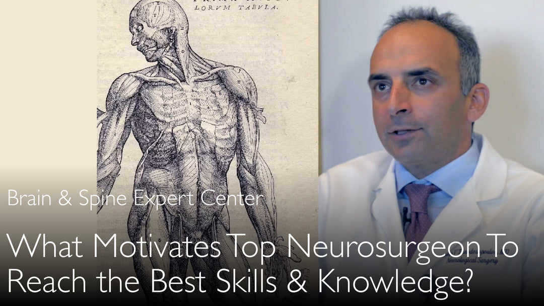
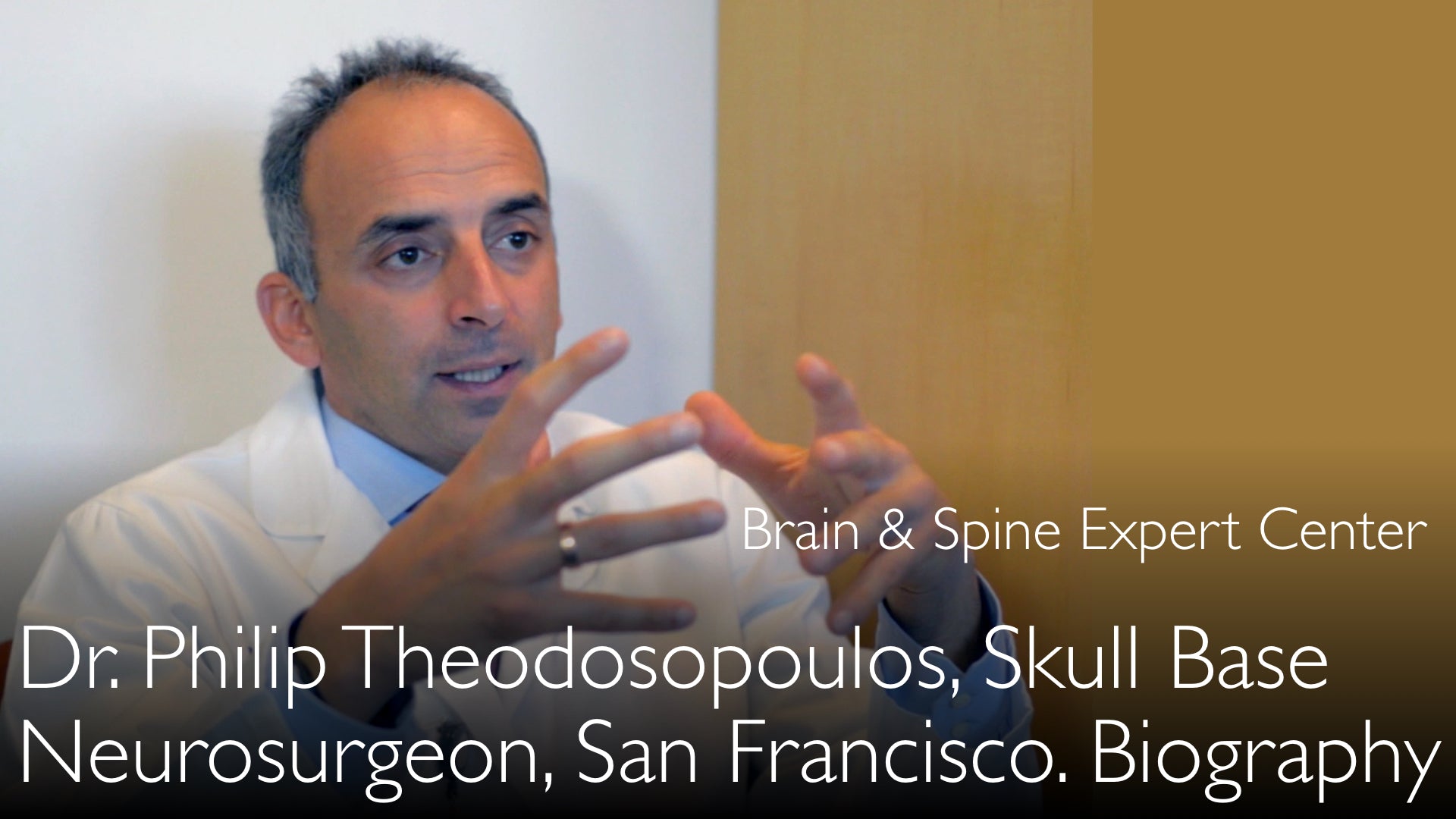
מומחה מוביל בנוירוכירורגיה של כלי הדם המוחיים ובסיס הגולגולת, ד"ר פיליפ תיאודוסופולוס, MD, מסביר את התפקיד הקריטי של לימוד אנטומי מתמשך להשגת מצוינות כירורגית. הוא מפרט את שיטות המחקר הקפדניות שלו באמצעות סריקות CT ונתיחות גופות כדי לשלוט באנטומיה המורכבת של בסיס הגולגולת. ד"ר פיליפ תיאודוסופולוס, MD, מדגיש שאנטומיה נורמלית יכולה לאתגר אפילו את המנתחים המנוסים ביותר. הוא תומך בחזרה לעקרונות אנטומיים יסודיים כדי להרחיב את גבולות המיומנות הכירורגית ולהבטיח את בטיחות המטופלים.
More from Brain and Spine
More from Diagnostic Detectives Network
שליטה באנטומיה כירורגית לניורוכירורגיה עליונה של בסיס הגולגולת
קפיצה לפרק
- אנטומיה: היסוד למצוינות כירורגית
- שיטות מחקר בחקר אנטומי
- מדוע האנטומיה הנורמלית מהווה אתגר כירורגי
- נתיחה בקדוורים לפיתוח מיומנות
- גישות אנדוסקופיות ונקודות מבט אנטומיות
- ההכרח בלמידה מתמשכת לשליטה כירורגית
- תמליל מלא
אנטומיה: היסוד למצוינות כירורגית
ד"ר פיליפ תיאודוסופולוס, MD, טוען שהבנה מעמיקה של האנטומיה המיקרוכירורגית היא אבן היסוד הבסיסית עבור כל נוירוכירורג השואף למצוינות ברמה עולמית. הוא מתחקה אחר שורשי הכירורגיה המודרנית עד לחלוצי הרנסנס כמו אנדריאס וסאליוס, שהדגישו את החשיבות הקריטית של נתיחה אנושית. פרספקטיבה היסטורית זו תומכת באמונתו שכירורגים חייבים להיות מושתתים על ידע אנטומי מעל לכל. ד"ר תיאודוסופולוס טוען שמיומנות טכנית בלבד אינה מספקת ללא הבנה יסודית זו.
שיטות מחקר בחקר אנטומי
ד"ר פיליפ תיאודוסופולוס, MD, משתמש בגישת מחקר רב-ממדית כדי לפרק את המורכבויות של בסיס הגולגולת. עבודתו הפורייה כוללת מחקרים קפדניים על גולגלות אנושיות יבשות באמצעות הדמיית טומוגרפיה ממוחשבת (CT) מתקדמת של 64-פרוסות. לדוגמה, מחקר אחד ניתח 84 גולגלות יבשות, בעוד אחר ביצע ניתוח מורפומטרי של 100 גולגלות באמצעות סריקות CT ומערכות ניווט Brain Lab.
הוא משלים ניתוח רדיולוגי זה עם נתיחות אנדוסקופיות ומיקרוכירורגיות מעשיות. מחקרו משתמש בראשי קדוורים ובדגימות מקובעים בפורמלין לתרגול וליטוש גישות כירורגיות, כמו אלה דרך הקונכייה האמצעית, הקונכייה התחתונה, והסינוס המקסילרי דרך גישת Caldwell-Luc.
מדוע האנטומיה הנורמלית מהווה אתגר כירורגי
עיקרון מרכזי בפילוסופיה של ד"ר תיאודוסופולוס הוא שהאנטומיה הנורמלית עצמה מהווה אתגר ראשוני בכירורגיה. הוא מספק אזהרה חדה: "אנטומיה נורמלית יכולה להפוך כירורג גדול לכירורג בינוני." במהלך ניתוחים, כירורגים נתקלים לרוב במבנים אנטומיים נורמליים, לא פתולוגיים. ללא ידע מעמיק ואינטימי של כל ניואנס ווריאציה שכיחה, ביצועי הכירורג יכולים להיפגע קשות.
זו הסיבה שלנתח חולים בלבד אינו מספיק לפיתוח מיומנות. ד"ר פיליפ תיאודוסופולוס, MD, מסביר לד"ר אנטון טיטוב, MD, שכירורג חייב לצאת מעבר לחדר הניתוח כדי ללמוד וללמוד מחדש באופן מתמשך את היחסים האנטומיים כדי להימנע מלהיות תת-אופטימלי.
נתיחה בקדוורים לפיתוח מיומנות
ד"ר פיליפ תיאודוסופולוס, MD, תומך בנתיחת קדוורים ככלי indispensable לחידוד מיומנויות כירורגיות וחדשנות בבטחה. הוא קובע שתרגול טכניקות חדשות בסביבה מדומה או על חומר קדוורי הוא הכרח אתי לפני ניסיונן על חולים. לקיחת טכניקות לא בדוקות ישירות לחולה מהווה "ניסוי בבני אדם", שבו הכירורג עשוי עדיין לא להיות מיומן.
שיטה זו מאפשרת לכירורגים לצבור ניסיון נרחב ללא סיכון לחולים. זו הדרך היחידה "לדחוף את הגבולות" של מה שאפשרי כירורגית בבטחה, להבין את המגבלות המדויקות של גישות שונות, ולהבין fully את הסיכונים הכרוכים בהליך.
גישות אנדוסקופיות ונקודות מבט אנטומיות
מוקד משמעותי בעבודה האחרונה של ד"ר תיאודוסופולוס הוא על שליטה באנטומיה עבור כירורגיה אנדוסקופית אנדונזלית של בסיס הגולגולת. הוא מציין שאנטומיה מוכרת, כמו בסיס הגולגולת, נראית完全不同 different כאשר ניגשים אליה מלמטה (אנדונזלית) compared to גישה גולגולתית מסורתית. בעוד שהאנטומיה עצמה אינה משתנה, נקודת המבט ומעברי הניתוח כן.
זה דורש לימוד ייעודי כדי להבין את המגבלות והיחסים של מבנים מנקודת תצפית ספציפית זו. מחקרו into areas like העורק הקרוטידי הפנימי, ה-optic strut, and fissure ארובתית תחתונה מנקודת מבט אנדוסקופית הוא crucial לפיתוח טכניקות minimally invasive בטוחות ויעילות.
ההכרח בלמידה מתמשכת לשליטה כירורגית
עבור ד"ר תיאודוסופולוס, החיפוש אחר ידע אנטומי אינו שלב בהכשרה אלא מחויבות לכל החיים. הוא מדגיש שלימוד rigorous ומתמשך זה הוא crucial not only להכשרת מתמחים וכירורגים עמיתים אלא ל"הכשרת עצמנו". זהו תהליך מתמשך שכל כירורג must engage in throughout הקריירה שלהם.
מסירות זו ללמידה מתמדת is what separates כירורגים טובים מכירורגים מובילים truly. ד"ר אנטון טיטוב, MD, מדגיש שרמת מסירות זו היא rare, comparing עבודת ד"ר תיאודוסופולוס ללכת בעקבות leonardo da vinci. מרדף relentless זה ensures שכירורג remains grounded, understands their limits, and ultimately provides the highest standard of care to their patients.
תמליל מלא
ד"ר אנטון טיטוב, MD: מה motivates כירורגים מובילים להצטיין בעבודה ובחיים? נוירוכירורג מוביל בכלי דם מוחיים ובבסיס הגולגולת משתף את התשוקה והמסירות שלו.
ד"ר פיליפ תיאודוסופולוס הולך בדרכו של אנדריאס וסאליוס ולאונרדו דה וינצ'י. פרופ' פיליפ תיאודוסופולוס לומד meticulously את המיקרואנטומיה הכירורגית של בסיס הגולגולת. He uses modern methods of CT scans to better understand every nuance of skull base anatomy.
"כירורגים mostly see normal anatomy when they operate on patients. Normal anatomy can reduce a great surgeon to a mediocre surgeon." "To be a superior surgeon, you have to go back to the basic building blocks of anatomy."
"In 84 dry human skulls, imaging studies were performed by 64-slice computed tomography." What motivates leading surgeons? ד"ר אנטון טיטוב, MD. What drives a highly skilled surgeon to continue studying the dissection anatomy of the skull? ד"ר פיליפ תיאודוסופולוס, MD.
A video interview with a leading expert in skull base neurosurgery. Microsurgical anatomy of the skull is necessary to improve surgical skill. Surgeons mostly see normal anatomy when they operate on patients. Normal anatomy can reduce a great surgeon to a mediocre surgeon.
A medical second opinion from a leading surgeon can confirm a tumor diagnosis. A medical second opinion also helps to choose the best treatment for cancer or a benign brain tumor. ד"ר אנטון טיטוב, MD. Seek a medical second opinion on a brain tumor and be confident that your treatment happens according to leading international standards.
ד"ר פיליפ תיאודוסופולוס, MD: Surgeons can only become better with more experience. But operating on patients is not sufficient. A surgeon has to continuously study dissection anatomy in his specialty. A neurosurgeon has to study brain anatomy on cadaveric heads.
To become a great surgeon, to push the envelope, a surgeon really has to go back to the basic building blocks of microsurgical dissection anatomy. What motivates leading surgeons? Becoming the best surgeon.
ד"ר אנטון טיטוב, MD: You have a particular interest in the delicate and very complex anatomy of the skull base. You prolifically publish this fascinating surgical anatomy research. Let me quote some titles from your six recent papers about the surgical anatomy of neurovascular skull base structures.
Endoscopic, endonasal variability in the anatomy of the internal carotid artery. Anatomic variation of the optic strut: classification schema, radiologic evaluation, surgical relevance. ד"ר אנטון טיטוב, MD. Anatomy of the inferior orbital fissure: implications for endoscopic cranial base surgery. Anatomic study of the prechiasmatic nucleus and its surgical implications.
ד"ר פיליפ תיאודוסופולוס, MD: Anatomy of the optic canal: a computed tomography study of endoscopic nerve decompression. Endoscopic anatomy of the petrous segment of the internal carotid artery.
Let me also quote some methods that you use in some of these studies: "In 84 dry human skulls, imaging studies were performed by 64-slice computed tomography." Endoscopic endonasal dissections were performed in six formalin-fixed cadaver heads.
Morphometric analysis of 100 skulls was conducted using CT scans and the Brain Lab. Four patients underwent procedures that exposed the maxillary strut. In 10 cadaveric specimens, microanatomical and endoscopic dissections were performed.
ד"ר פיליפ תיאודוסופולוס, MD: Dissections were done via approaches in the middle turbinate and the inferior turbinate. Dissections were performed also via the Caldwell-Luc treatment through the maxillary sinus.
These quotes from your work and your studies vividly demonstrate the degree of dedication. They show the need for continuous honing of surgical skills. They also show your understanding of very specific and delicate surgical anatomy.
ד"ר פיליפ תיאודוסופולוס, MD: Such technical skills are required for a surgeon who wishes to have world-level excellence in his field. Not many surgeons today are literally following the steps of Leonardo da Vinci. Not many surgeons do their own anatomical research today.
ד"ר אנטון טיטוב, MD: What drives you in these rigorous pursuits? How does it help you in your clinical practice?
ד"ר פיליפ תיאודוסופולוס, MD: It is interesting. Microsurgical anatomy has been my primary interest in research. My other interest is clinical outcomes research.
Medicine may have started with Hippocrates and Galen. But surgery really started with Andreas Vesalius and patients in the Middle Ages and Renaissance. In the Renaissance, they actually took an interest in dissecting the human body.
It is the only method how surgeons can really stay grounded in what is important in surgery. We need understanding that even the most experienced surgeons are reduced to mediocre surgeons so often by the normal anatomy that we encounter.
It goes without saying that for a surgeon to be good, a surgeon has to really study a lot.
ד"ר אנטון טיטוב, MD: The studying doesn't come only from the patients you see. Because there is just no method that you can see this many patients or do this many surgeries. To some degree, it ends up being human experimentation.
Sometimes you are not good at what you are devising. All of these new techniques that we have.
ד"ר פיליפ תיאודוסופולוס, MD: Sometimes you take the new techniques straight to the patient instead of having done that many, many times in cadaveric material. Or you practiced new techniques in a simulated environment.
You are not able to say that we are safe to perform a new surgical technique on a patient. That is what we see in this anatomic work. We see that we can only become better surgeons by experience.
If you want to push the envelope, you really have to go back to basics of surgical methods. You have to go back to the basic building blocks of anatomy. You have to understand anatomy very well.
עליכם להבין את הקשרים בין המבנים האנטומיים. העבודה העדכנית ביותר שביצענו מתמקדת בשיטות האנדוסקופיות לטיפול בבסיס הגולגולת.
בסיס הגולגולת שראינו אלפי פעמים מהגישה הקדמית. בסיס הגולגולת שאנו רואים מהאזור התת-גולגולתי. הוא נראה כל כך שונה כשמגיעים מלמטה. זו אותה אנטומיה.
האנטומיה אינה משתנה בין אם השיטה שלנו מגיעה מלמטה, מהגישה הקדמית, או מהצד. עליכם ללמוד ולהבין את המגבלות, הקשרים, וכיוצא בזה, מהטיפול הספציפי שאתם מבצעים.
רק אז לא תהיו טובים. לא תבינו את הגבולות. לא תבינו את הסיכונים שאתם נוטלים. אז תהיו תת-מיטביים בכישוריכם הכירורגיים.
בהכשרה, זה קריטי עבור כולנו. זה לא רק קריטי בהכשרת המתמחים או העמיתים הכירורגיים שלנו.
Dr. Anton Titov, MD: אלא בהכשרת עצמנו, בהכשרת כולנו, זה הכרחי שעלינו לעשות זאת. עלינו ללמוד אנטומיה כירורגית ברציפות ובקפדנות.
מה מניע כירורגים מובילים? כיצד להשיג כישורים כירורגיים מובילים? ראיון וידאו עם מומחה מוביל בנוירוכירורגיית בסיס הגולגולת. מצוינות כירורגית ואנטומיה.