מומחה מוביל בנוירופתולוגיה, ד"ר סבסטיאן ברנדנר, MD, מסביר כיצד אבחון מולקולרי מחולל מהפכה באבחון ובטיפול בגידולי מוח. הוא מפרט את התפקיד הקריטי של בדיקת מוטציית IDH בגליומות ובאסטרוציטומות, תוך הדגשת המקרים שבהם היסטולוגיה מסורתית נותרת מספקת לאבחון מנינגיומה.


אבחון מולקולרי בפתולוגיה של גידולי מוח: ממנינגיומה לגליובלסטומה
קפיצה לפרק
- היסטולוגיה מסורתית באבחון גידולי מוח
- אבחון פשוט של מנינגיומות שפירות
- בדיקה מולקולרית לגליומות ולמוטציות IDH
- סמנים ביולוגיים מרכזיים באבחון אסטרוציטומה
- גורמי פרוגנוזה מעבר לסוג הגידול
- עתיד האבחון של גידולי מוח
- תמליל מלא
היסטולוגיה מסורתית באבחון גידולי מוח
ד"ר סבסטיאן ברנדנר, MD, מדגיש שהיסטולוגיה בסיסית נותרת הבסיס של פתולוגיית גידולי מוח. שקופית הצביעה הראשונית H&E (המטוקסילין ואאוזין) מספקת תובנות ראשוניות קריטיות, כשדגימות רקמה מוכתמות בוורוד חושפות את ארכיטקטורת הגידול תחת המיקרוסקופ. שיטה מסורתית זו ממשיכה להנחות את מסלול האבחון במקרים רבים של גידולי מוח.
אבחון פשוט של מנינגיומות שפירות
מנינגיומות הנובעות מקרומי המוח לרוב אינן דורשות בדיקה מולקולרית, כפי שמסביר ד"ר סבסטיאן ברנדנר, MD. גידולים שפירים אלו typically can be definitively diagnosed through standard histology alone. הנוירופתולוג מציין שמיקום הגידול - במיוחד positions at the base of the skull that are difficult to access - often impacts prognosis more than molecular factors in meningiomas. מנינגיומות שטחיות generally have excellent outcomes with complete surgical resection.
בדיקה מולקולרית לגליומות ולמוטציות IDH
בגליומות, especially in younger patients, ד"ר סבסטיאן ברנדנר, MD, highlights the critical importance of IDH mutation testing. A specialized antibody developed at the German Cancer Research Center detects 90% of IDH1 mutations and rare IDH2 variants. This cost-effective immunohistochemical method provides rapid, reliable results that significantly influence treatment decisions and prognosis assessment.
סמנים ביולוגיים מרכזיים באבחון אסטרוציטומה
ד"ר ברנדנר מתאר גישת two-antibody approach for astrocytoma diagnosis. The IDH mutation test combines with analysis of nuclear protein loss - when the characteristic "black dot" disappears from cell nuclei, it strongly indicates astrocytoma. This molecular signature helps differentiate astrocytomas from other glioma subtypes and guides appropriate treatment strategies.
גורמי פרוגנוזה מעבר לסוג הגידול
While molecular diagnostics provide essential information, ד"ר ברנדנר stresses that tumor location and surgical accessibility remain crucial prognostic factors. Even benign tumors in challenging anatomical positions may have poorer outcomes than more aggressive tumors in operable locations. הנוירופתולוג emphasizes the need for comprehensive evaluation combining histological, molecular and clinical data.
עתיד האבחון של גידולי מוח
ד"ר סבסטיאן ברנדנר, MD, anticipates continued advancement in molecular pathology techniques. The success of IDH mutation testing demonstrates how targeted biomarkers can streamline diagnosis while improving accuracy. As research identifies more tumor-specific molecular signatures, pathology laboratories will increasingly combine traditional and molecular methods for optimal patient care.
תמליל מלא
ד"ר אנטון טיטוב, MD: בעבר, גידולי מוח אובחנו באופן relatively crude. Basic staining is still the first step in the pathology analysis of brain cancer. But now there is more molecular diagnostics and molecular analysis of brain tumor mutations. This becomes very important for treatment of tumors. It is important for the brain tumor prognosis.
ד"ר אנטון טיטוב, MD: What is the importance of molecular diagnostics for brain tumor diagnosis and therapy?
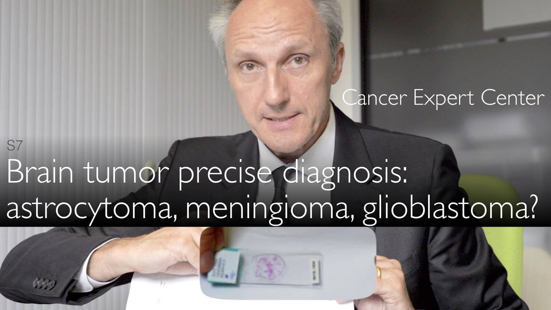
ד"ר סבסטיאן ברנדנר, MD: First of all, you are absolutely correct that the mainstay of pathology diagnostics is still the first histology slide. It is a slide that looks like this. It has pink staining. I just give an example. This is the size of a slide - 1 by 3 inches. Sometimes you can see against the background of these little pink flics in the center. These are the brain tumor tissue specimen.
ד"ר סבסטיאן ברנדנר, MD: We put this slide first under the microscope. That is the first decision in brain tumor diagnosis.
ד"ר אנטון טיטוב, MD: What to do next?
ד"ר סבסטיאן ברנדנר, MD: First of all, there might be a number of tumors, like the benign meningioma. Sometimes a brain tumor arises from the coverings of the brain rather than from within the brain. Meningiomas are usually diagnosed just on this section slide. So it's a fast and cheap brain tumor diagnosis. It gives the best information.
ד"ר סבסטיאן ברנדנר, MD: There is no need to do any further molecular testing. Because there are other factors determining whether this brain tumor will have bad prognosis or a good prognosis. For example, where the brain tumor grows. Sometimes it grows in difficult to reach areas. Bottom of the brain, which is called "skull base". Then this determines the clinical prognosis.
ד"ר סבסטיאן ברנדנר, MD: The brain tumor that is easily accessible on the surface of the brain can be taken out. Usually these brain tumors don't recur. If meningiomas recur, they can often be resected a second time.
ד"ר סבסטיאן ברנדנר, MD: These are the meningiomas. You can diagnose them just on single H&E slide. Gliomas generally can also be diagnosed on a single H&E simple staining slide, particularly glioblastomas.
ד"ר סבסטיאן ברנדנר, MD: But there are patients who are younger. Some patients have gliomas that have mutations that I mentioned earlier. Gliomas have IDH mutation. These IDH mutations can be detected with an antibody. This antibody was generated in the laboratory in Heidelberg at the German Cancer Research Center. This antibody detects the glioma mutation. So it's a cheap and quick, pathologist-friendly way to diagnose mutations in a brain tumor.
ד"ר סבסטיאן ברנדנר, MD: The nice thing is that this antibody detects 90% of all IDH mutations. 95% of IDH1 mutations and very rare, so-called IDH2 mutations in glioma. This antibody is then combined with another antibody. That antibody detects a nuclear protein. It should normally be present in the cell in the nucleus.
ד"ר סבסטיאן ברנדנר, MD: But in some type of brain tumors this protein is lost in the nucleus. These brain tumors are astrocytomas. So the small black dot is lost from the cell's nucleus. We would normally find this black dot in the middle of the cell, in the nucleus. That loss is characteristic and nearly diagnostic for astrocytoma.
ד"ר אנטון טיטוב, MD: IDH mutation is present also in astrocytoma brain tumor. Brain tumor diagnosis expert explains precise molecular diagnosis of brain tumor type. Benign meningioma. More aggressive astrocytoma and glioblastoma multiforme (GBM).